The group is involved in a wide range of projects in a number of areas.
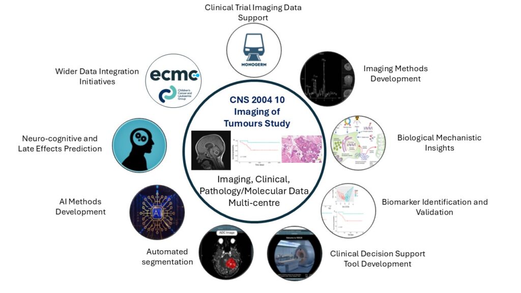
These projects include:
- Clinical Trial Imaging Data Support
- Imaging Methods Development
- Biological Mechanistic Insights
- Biomarker Identification and Validation
- Clinical Decision Support Tool Development
- Automated Segmentation
- AI Methods Development
- Neuro-cognitive and Late Effects Prediction
- Wider Data Integration Initiatives
Ongoing/Current Projects
- Developing a clinical decision support tool for non-invasive diagnosis of childhood cancers
- Advancing radiomic tools for rare brain tumours: Enriching data sets and developing novel AI approaches. Funded by Great Ormond Street Biomedical Research Centre
- Metabolic and Imaging Characteristics of Senescence in Childhood Brain Tumours: Clinical PhD Student ship from the Azaylia Foundation
Group member Dr Katie Crombie, one of the Azaylia PhD fellows took part in two videos for the foundation. Take a look at a Week in the life of a PhD researcher and learn more about Katie’s PhD. - Automatic Segmentation of Pediatric Brain Tumours using Diffusion-weighted MRI: Downstream Evaluation on Diagnostic Classification Task – Dr Jan Novak, Aston University
- Assessing variable survival in paediatric brain tumours – Dr James Grist, Oxford University
- Mapping brain microstructural damage in paediatric posterior fossa tumour survivors, Prof Chris Clark, Great Ormond Street Hospital.
Developing and understanding new treatments for craniopharyngioma - Dr John Apps

Craniopharyngioma is a type of childhood brain tumour that is difficult to treat. It is in an area of the brain that makes the tumours hard to remove with surgery, and radiotherapy doesn’t work fully in a quarter of children. New treatments are needed to improve the future for these children.

Dr John Apps and his team at the University of Birmingham have found a group of medicines that could help stop tumours from growing. They are called MEK inhibitors and IL6 inhibitors, and will be tested in an international clinical trial for regrown (relapsed) craniopharyngiomas. The research team will be analysing tumour samples from the trial to find out more about what goes on inside the tumour and why they relapse. They will also look at the large cysts that often occur when craniopharyngiomas regrow to find out what they are and what treatments could help.
Archive of previous projects
Lab based research
‘The role of glutamate metabolism in Medulloblastoma’ Haydn Munford
I am looking at the effect of a particular chemical in the cell called glutamine, to find out if it can help make cancer treatments work better.
‘The role of lipid droplets in glioblastoma’ Rob Murren
The focus of my project is to investigate the role of fat droplets in glioblastoma and understand how we may alter them to improve current treatments.
Imaging
‘The development of a novel MRI based method for measuring blood perfusion in neurovascular damage’ Emma Metcalfe-Smith
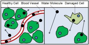
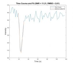
My work focuses on developing a new way to measure the movement of blood and water within body tissue using MRI (Magnetic Resonance Imaging). A greater understanding of this movement will help us to better understand the behaviour of different tissues. It is hoped that this will give more accurate measurements which will help in the diagnosis and treatment of injuries such as damage to nerves and blood vessels.
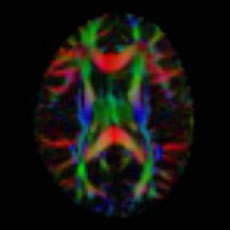
‘The Role of Diffusion Tensor Imaging in the Diagnosis of Paediatric Brain Tumours’ Heather Rose
We focus on the use of advanced magnetic resonance imaging (MRI) to help diagnose brain tumours in young patients. We measure the movement of water molecules within the brain to help identify any change in structure in and around brain tumours. An additional focus of the research project is the design of databases that store the MRI images and allow researchers from different universities to work on joint research projects.
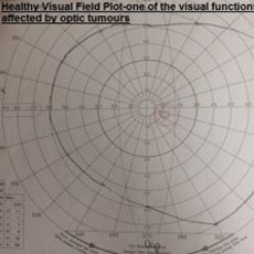
‘The Fusion of MRI and Diffusion Imaging (DTI) of the optic pathway as an aid to the management of Optic Pathway Glioma (OPG)’ Lara Worthington
My research is aimed at trying to use MRI (Magnetic Resonance Imaging) to indicate possible damage to children’s vision arising from tumours located near the optical nerves in the brain.
‘Communicating information from MRI – Patient and parent views’ Natalie Tyldesley-Marshall
I am interviewing young patients and their parents about how they feel about, understand and value seeing the MRI (Magnetic Resonance Imaging) images of their / child’s brain tumour. We hope this will help better guide discussions between doctors and patients around their test results.
‘Measuring blood flow in childhood brain tumours using magnetic resonance imaging’ Stephanie Withey
My research involves measuring blood flow in childhood brain tumours using magnetic resonance imaging (MRI). While patients are having their scan they are given an injection of a dye which enters the brain tumour and changes how it looks on the pictures from the MRI. I write computer programs that use these changes to give us extra information on the type of tumour it is and how well the treatment is working.
‘Development of novel methods for obtaining robust biomarkers from diseased and ageing brains’ Stephen Powell
My PhD focuses on developing methods to automatically assess image quality and obtain biomarkers from MRI (Magnetic Resonance Imaging). Biomarkers are quantities which can be measured and used to determine whether the brain is healthy or not.
‘The cutting-edge research of the advanced MR imaging technology in biomedical and brain science’ Yu Sun
My research focuses on using MRI (Magnetic Resonance Imaging) to better understand tumours in children and how to identify these earlier. My project looks at how to use the latest A.I. (Artificial Intelligence) to investigate brain disease and improve chances of survival.
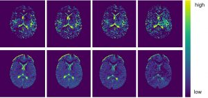
‘Investigation of Tissue Microenvironments using Diffusion Magnetic Resonance Imaging’ Emma Meeus
In my project we hope to improve the measurement of diffusion with magnetic resonance imaging (MRI) and the following image analysis methods. This aims to provide a quicker way to identify and assess the characteristics of childhood tumours.
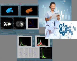
‘Developing a clinical decision support system using the latest diagnostic analysis techniques’ Niloufar Zarinabad
My current project is to translate the latest MRI (Magnetic Resonance Imaging) and latest methods towards practice in the clinic. We are designing a new system that will help doctors make decisions by providing new information and decide on which treatments are best for patients and improve their care.
Spectroscopy
‘Utilising non-invasive MRI measures to predict survival outcomes in childhood brain tumour patients‘ Ben Babourina-Brooks
In my research I use advanced MRI (Magnetic Resonance Imaging) techniques to measure chemicals and temperature in the brain. I use these techniques to help us distinguish between different types of tumours to help the clinic make better treatment decisions for patients.
‘Advanced MRI of childhood brain tumours’ Chris Bennett
My work involves working on brain tumour tissue frozen after surgery. This allows us to examine real tumour tissue in more detail than we can with an MRI (Magnetic Resonance Imaging) scanner, helping us to better understand imaging and spectroscopic features of brain tumours.
‘Diagnosing brain tumour through functional imaging and deep learning’ Dadi Zhao
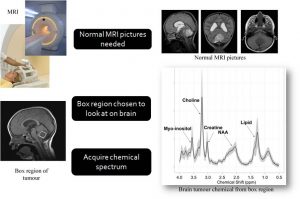
Conventional MR (magnetic resonance) imaging is able to provide useful information on the structure, and functional imaging (spectroscopy) can aid getting estimates of certain chemicals (called metabolites) in the tumour. In this project I am trying to find a novel way to realise automatic brain tumour classification through machine learning, in the same exact area of the brain, to better determine which type and grade of brain tumour a patient has.
‘Establishing the added value of advanced imaging techniques in clinical practice’ Karen Manias
My research involves establishing how the newest MRI (Magnetic Resonance Imaging) methods help doctors diagnose children with cancer and better understand how well the treatment is working. the main aim of my work is to take the latest imaging methods developed by the CBTRT back to the doctors in the clinic to improve patient care.
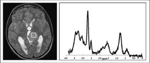
‘Non invasive identification of medulloblastoma genetic subtypes using metabolite profiles and imaging features’ Sarah Kohe
My main project is funded by Children with Cancer UK and involves the development of rapid and non-invasive methods of identifying the different genetic types of medulloblastoma based on their metabolite (chemical) profiles and the other features that we can see using MRI (Magnetic Resonance Imaging). Finding out how we can diagnose (or identify) these earlier and non-invasively will help doctors better plan treatment, and have more informed discussions with the patient and their family in the crucial early stages after the tumour has been found. Improving our understanding of the metabolite pathways in these different types (or subgroups) of medulloblastoma also means that we might find new ways to treat this.